CASK gene disorders
Diagnosis and testing
Find out more about how the conditions are diagnosed.

Genetic testing
If your child has symptoms that could be due to a CASK gene mutation, such as microcephaly, their DNA will be tested. The test looks for changes in the gene that cause a CASK-related disorder.
Sometimes parents receive this information over the phone, which can be difficult. If you need to speak to someone about your results, please contact us. It's important to obtain a copy of the test results, which we can then go through with you as needed.
Testing for different mutations
To understand the different mutations and why they can cause different symptoms, read our CASK Gene for Beginners page.
Missense, nonsense and frameshift mutations are usually detected by gene panels, whole exome sequencing (WES) or whole genome sequencing (WGS). Splice site mutations can also be detected using any of these three techniques, but sometimes additional RNA testing may help clarify their effect.
Deletions and duplications may be harder to detect. They are often found using chromosomal microarray, MLPA testing, whole genome sequencing (WGS), or some exome tests with copy number analysis.
Detecting mosaicism can be difficult, especially if only a small percentage of cells carry the mutation. Higher-depth sequencing or testing different tissues may occasionally be needed to detect this form of the disease.
We are members of Genetic Alliance UK, who create resources for families. The Rare Resources guides are produced for families who have recently received a diagnosis, are on the journey to a diagnosis, or who have been told their child's condition is so rare they might not get a diagnosis. Download the guide.
Understanding your genetics report
Genetic reports are difficult to interpret unless you're a geneticist or genetic counsellor. We regularly have parents asking us for help. As a result we have created a plain-English guide to assist families.
Read the guide →A wide spectrum of conditions
CASK-related disorders exist on a broad spectrum. Even people with the same type of mutation can be affected differently.
Researchers are still learning why some mutations cause severe MICPCH while others lead to milder XL-ID presentations. Factors such as mosaicism, X-chromosome inactivation, and how much working protein remains may all play a role.
As understanding of CASK continues to grow, improved genetic testing is helping more families receive accurate diagnoses and better information about their child's condition.

Diagnosing MICPCH
Many females with MICPCH are first diagnosed with microcephaly and developmental delay, then receive an MRI to image the brain. A diagnosis of PCH (pontocerebellar hypoplasia) may be given to families, who will await genetic results to confirm the genetic cause of the PCH.
In the UK it can take six months to receive the genetic test results to confirm a CASK mutation. The radiological features below help distinguish between some forms of PCH and CASK-related MICPCH, enabling families to get a more definitive answer faster. Any diagnosis should always be followed up by a genetic test to confirm a CASK mutation.
Distinguishing CASK-related MICPCH from PCH type 2 and 4
Although CASK is the most frequently mutated gene associated with PCH, it is one of the least well-known and is often overlooked.
The majority of female patients with a mutation in their CASK gene have an underdevelopment of the cerebellum, the cerebellar vermis, and the pons, called pontocerebellar hypoplasia (PCH). Usually there is a proportionate involvement of the cerebellar vermis and hemispheres, contrary to PCH type 2 and type 4 (Moog et al. 2011; ref). These types of PCH are predominantly associated with flat cerebellar hemispheres and a small but less severely affected vermis, giving a dragonfly-like appearance to the cerebellum.
In female patients with a CASK mutation the corpus callosum tends to have a normal size. Since the corpus callosum is normal in size and the cerebrum is smaller (because of the microcephaly), this can give the impression of thickening of the corpus callosum (Takanashi 2010).
In addition to brain imaging results, specific clinical features (or their absence) can help differentiate a CASK-related disorder from PCH type 2. Generalized clonus ("jitteriness"), lack of voluntary motor development, spasticity, impaired swallowing, and epilepsy shortly after birth are commonly associated with PCH2 but are rarer or absent in female patients with CASK mutations, whilst sensorineural hearing loss may be suggestive of CASK.
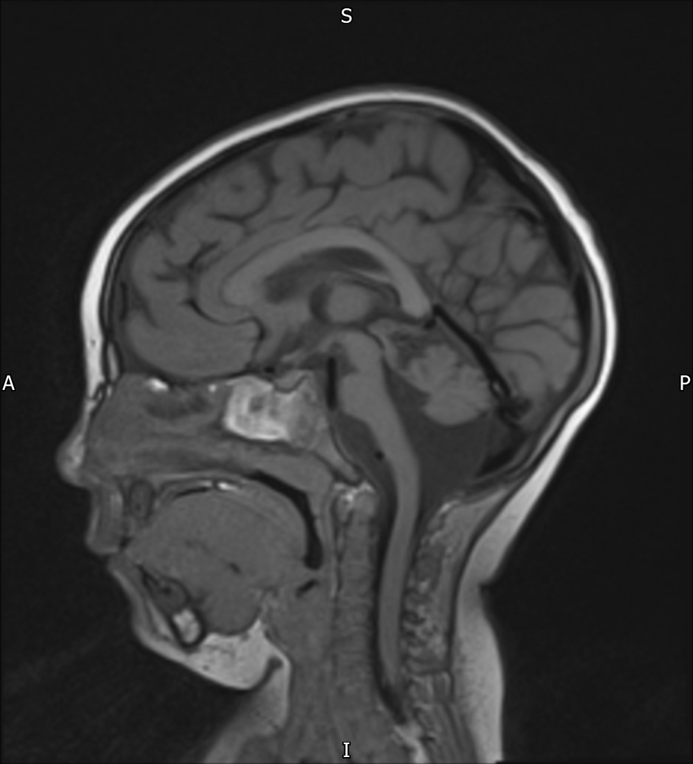
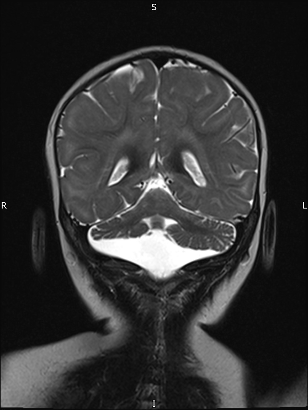
MRI images of a patient with a CASK mutation displaying typical MICPCH features.
This information was created by CASK Research in partnership with Professor Kutsche and Dr Amin.